Heart Anatomy⁚ A Comprehensive Overview
The heart is a vital organ responsible for pumping blood throughout the body, delivering oxygen and nutrients to tissues and removing waste products. Understanding the intricate anatomy of the heart is crucial for comprehending its function and for diagnosing and treating cardiovascular diseases. This comprehensive overview will delve into the various components of the heart, exploring its structure, chambers, valves, blood flow, and associated structures.
Introduction
The heart, a remarkable muscular organ, lies at the very core of the circulatory system, serving as the tireless pump that propels life-sustaining blood throughout the body. Its intricate anatomy, a symphony of chambers, valves, and specialized tissues, orchestrates the rhythmic flow of blood, delivering oxygen and vital nutrients while removing waste products. This intricate dance of blood flow is essential for maintaining every function of the body, from the beating of the brain to the contraction of muscles.
The study of heart anatomy is fundamental to understanding the complexities of cardiovascular health. By delving into the structure and function of this vital organ, healthcare professionals gain invaluable insights into the causes and mechanisms of cardiovascular diseases, paving the way for effective diagnosis, treatment, and prevention.
This exploration of heart anatomy will provide a comprehensive overview of the heart’s structure, its chambers, the intricate network of valves that regulate blood flow, and the coronary circulation that nourishes the heart itself. We will also delve into the unique properties of cardiac muscle, the protective pericardium, and the electrical conduction system that orchestrates the rhythmic beating of the heart.
By understanding the intricacies of heart anatomy, we gain a deeper appreciation for the remarkable resilience and complexity of this essential organ, paving the way for a greater understanding of its health and well-being.
Location and Structure
The heart, a vital organ responsible for circulating blood throughout the body, resides within the thoracic cavity, nestled in the mediastinum, the space between the lungs. It is positioned slightly to the left of the midline, with its apex (pointed end) pointing downwards towards the left side of the body. This strategic location provides the heart with optimal protection while allowing for efficient blood flow to all regions of the body.
The heart is a complex and fascinating structure, resembling a cone-shaped muscular organ. Its external surface is characterized by a series of grooves, known as sulci, which mark the boundaries between the heart’s four chambers. The coronary sulcus, the most prominent groove, separates the atria (upper chambers) from the ventricles (lower chambers). The interventricular sulci, located on the anterior and posterior surfaces of the heart, mark the division between the right and left ventricles.
The heart’s structure is further defined by its walls, which are composed of three distinct layers. The epicardium, the outermost layer, is a thin, protective membrane that also houses the coronary arteries and veins. The myocardium, the middle layer, is the thick, muscular wall responsible for the heart’s powerful contractions. The endocardium, the innermost layer, lines the chambers of the heart and is composed of smooth, endothelial cells that facilitate smooth blood flow.
Chambers of the Heart
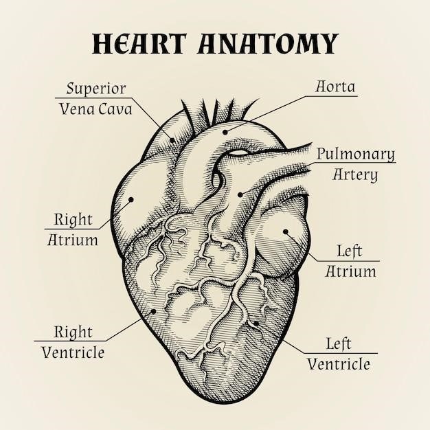
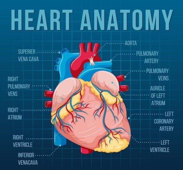
The heart is a four-chambered organ, each chamber playing a crucial role in the circulatory system. The two upper chambers, the atria, receive blood from the body and the lungs. The right atrium receives deoxygenated blood from the body through the superior and inferior vena cava, while the left atrium receives oxygenated blood from the lungs through the pulmonary veins. The two lower chambers, the ventricles, are responsible for pumping blood out of the heart. The right ventricle pumps deoxygenated blood to the lungs through the pulmonary artery, while the left ventricle pumps oxygenated blood to the body through the aorta, the largest artery in the body.
The right atrium and right ventricle work together to form the right side of the heart, which handles deoxygenated blood. The left atrium and left ventricle form the left side of the heart, which handles oxygenated blood. This separation ensures that oxygenated and deoxygenated blood do not mix, maintaining efficient oxygen delivery to the tissues. The chambers of the heart are separated by valves that regulate blood flow, ensuring it moves in the correct direction and prevents backflow.
The right and left atria are separated by the interatrial septum, a thin wall that contains the foramen ovale in the fetal heart. The right and left ventricles are separated by the interventricular septum, a thick muscular wall that forms the floor of the heart. These septa are essential for maintaining the separation between the oxygenated and deoxygenated blood circuits.
Valves of the Heart
The heart’s valves are crucial for maintaining unidirectional blood flow, preventing backflow, and ensuring efficient circulation. These valves act like one-way gates, opening to allow blood to flow in one direction and closing to prevent it from flowing back. The heart contains four valves⁚ the tricuspid valve, the pulmonary valve, the mitral valve, and the aortic valve.
The tricuspid valve, located between the right atrium and the right ventricle, prevents backflow of blood from the ventricle into the atrium during ventricular contraction. The pulmonary valve, situated at the opening of the pulmonary artery, prevents backflow of blood from the pulmonary artery into the right ventricle during ventricular relaxation.
The mitral valve, also known as the bicuspid valve, is located between the left atrium and the left ventricle. It prevents backflow of blood from the ventricle into the atrium during ventricular contraction. The aortic valve, positioned at the opening of the aorta, prevents backflow of blood from the aorta into the left ventricle during ventricular relaxation.
The valves are composed of thin flaps of tissue called cusps, which are attached to the walls of the heart by chordae tendineae, tiny, strong, fibrous strings. These strings are connected to papillary muscles, which are projections of the ventricular walls. This intricate structure ensures that the valves open and close properly, preventing backflow and maintaining efficient blood circulation.
Blood Flow Through the Heart
The heart is a complex pump that drives the continuous circulation of blood throughout the body. This circulatory system is responsible for delivering oxygen and nutrients to the tissues and removing carbon dioxide and waste products. The flow of blood through the heart follows a specific pathway, ensuring that deoxygenated blood is oxygenated and then distributed to the body.
The journey begins with deoxygenated blood returning from the body through the superior and inferior vena cava, entering the right atrium. From there, it flows through the tricuspid valve into the right ventricle. The right ventricle pumps the blood through the pulmonary valve into the pulmonary artery, which carries it to the lungs for oxygenation.
In the lungs, carbon dioxide is released and oxygen is absorbed. The now oxygenated blood returns to the heart through the pulmonary veins, entering the left atrium. From the left atrium, blood flows through the mitral valve into the left ventricle. The left ventricle, the strongest chamber of the heart, pumps the oxygenated blood through the aortic valve into the aorta, the main artery that distributes blood to the rest of the body.
This continuous cycle of blood flow through the heart ensures that all tissues receive the oxygen and nutrients they need to function properly, while waste products are removed. The heart’s intricate structure and synchronized contractions enable this vital process to occur seamlessly, maintaining life.
Coronary Circulation
While the heart diligently pumps blood to the rest of the body, it also needs its own supply of oxygen and nutrients to function. This vital task is fulfilled by the coronary circulation, a network of blood vessels that specifically serve the heart muscle itself.
The coronary arteries, branching off from the aorta just above the aortic valve, provide the heart muscle with oxygenated blood. The left coronary artery, the larger of the two, divides into the left anterior descending artery (LAD) and the circumflex artery. The LAD supplies blood to the front and bottom of the left ventricle, while the circumflex artery supplies the left ventricle’s side and back.
The right coronary artery supplies blood to the right ventricle, the right atrium, and a portion of the left ventricle’s back. These arteries branch into smaller vessels, forming a vast network that reaches every part of the heart muscle.
Deoxygenated blood from the heart muscle is collected by the coronary veins, which eventually drain into the coronary sinus, a large vein that empties into the right atrium. This closed loop of coronary circulation ensures a constant supply of oxygen and nutrients to the heart, enabling its tireless work of pumping blood throughout the body.
Cardiac Muscle
The heart, a tireless pump, is composed of a unique type of muscle tissue known as cardiac muscle. This specialized tissue is responsible for the rhythmic contractions that propel blood throughout the body. Unlike skeletal muscle, which is under voluntary control, cardiac muscle is involuntary, meaning its contractions are controlled by the autonomic nervous system and the heart’s own internal pacemaker.
Cardiac muscle fibers are branched and interconnected, forming a complex network. This interconnectedness allows for coordinated contractions, ensuring that the heart beats as a single unit. The cells are also rich in mitochondria, the powerhouses of cells, which provide the energy needed for the heart’s continuous activity.
Cardiac muscle possesses a unique property called automaticity, meaning it can generate its own electrical impulses. This allows the heart to beat independently of the nervous system. The heart’s rhythm is regulated by a specialized group of cells known as the sinoatrial (SA) node, which acts as the heart’s natural pacemaker.
The structure and function of cardiac muscle are critical for the heart’s ability to pump blood effectively. Its unique characteristics, including its involuntary nature, interconnectedness, and automaticity, make it a vital component of the cardiovascular system.
Pericardium
The pericardium is a protective sac that encloses the heart, providing both support and protection. It is composed of two layers⁚ the fibrous pericardium and the serous pericardium. The fibrous pericardium, the outermost layer, is a tough, fibrous sac that anchors the heart to surrounding structures, preventing it from shifting excessively. It also helps to prevent overfilling of the heart with blood.
The inner layer, the serous pericardium, is composed of two sublayers⁚ the parietal pericardium, which lines the fibrous pericardium, and the visceral pericardium, which adheres directly to the heart’s surface. Between these two layers lies the pericardial cavity, a space filled with a small amount of lubricating fluid. This fluid reduces friction between the heart and the pericardium as the heart beats, allowing for smooth and efficient movement.
The pericardium plays a crucial role in maintaining the heart’s position within the chest cavity, preventing excessive stretching, and protecting it from infection. It also helps to maintain a constant pressure within the heart, which is essential for optimal functioning. In some cases, inflammation of the pericardium, known as pericarditis, can occur, leading to chest pain and other symptoms.
The pericardium, a seemingly simple sac, plays a vital role in the heart’s well-being, ensuring its smooth and efficient operation within the chest cavity.
Cardiac Conduction System
The cardiac conduction system is a specialized network of electrical cells within the heart that controls the heart’s rhythm and ensures coordinated contraction of the chambers. This intricate system acts as a sophisticated electrical network, generating and transmitting electrical impulses that regulate the heart’s beating. The system comprises several key components⁚
The sinoatrial (SA) node, often referred to as the “pacemaker” of the heart, is located in the right atrial wall. It spontaneously generates electrical impulses at a regular rate, initiating the heartbeat. From the SA node, the electrical signal travels to the atrioventricular (AV) node, situated at the junction of the atria and ventricles. The AV node acts as a gatekeeper, delaying the signal slightly to allow the atria to fully contract and fill the ventricles with blood before ventricular contraction begins.
The signal then travels through the bundle of His, a specialized pathway that divides into left and right bundle branches, extending down the interventricular septum. These branches further divide into Purkinje fibers, a network of fine fibers that spread throughout the ventricular walls. The Purkinje fibers rapidly transmit the electrical signal to the ventricular muscle cells, triggering their contraction and pumping blood out of the heart.
The coordinated activity of the cardiac conduction system ensures efficient and rhythmic pumping of blood, maintaining adequate blood flow throughout the body. Disruptions in this system can lead to arrhythmias, irregular heart rhythms, which can have serious consequences if left untreated.
Imaging Techniques for Heart Anatomy
Modern medical imaging techniques have revolutionized our understanding of heart anatomy, providing detailed insights into the structure and function of this vital organ. These non-invasive techniques allow healthcare professionals to visualize the heart in three dimensions, assess its chambers, valves, and blood flow patterns, and diagnose a wide range of cardiovascular conditions.
Echocardiography, often referred to as an “ultrasound of the heart,” uses sound waves to create images of the heart’s structure and function. It is a safe and widely used technique for assessing heart valve function, chamber size, and blood flow patterns.
Cardiac computed tomography (CT) scans use X-rays to create detailed images of the heart, revealing its structure, coronary arteries, and surrounding tissues. CT scans can detect blockages in the coronary arteries, identify abnormalities in the heart’s structure, and assess the effectiveness of heart procedures.
Magnetic resonance imaging (MRI) uses strong magnetic fields and radio waves to create detailed images of the heart, providing information about the heart’s structure, function, and blood flow. It is particularly useful for assessing heart muscle function, detecting congenital heart defects, and evaluating the effectiveness of heart treatments.

These imaging techniques have significantly advanced our ability to diagnose and treat heart conditions, enabling healthcare professionals to provide more precise and personalized care to patients.


